
The first 3D color X-rays
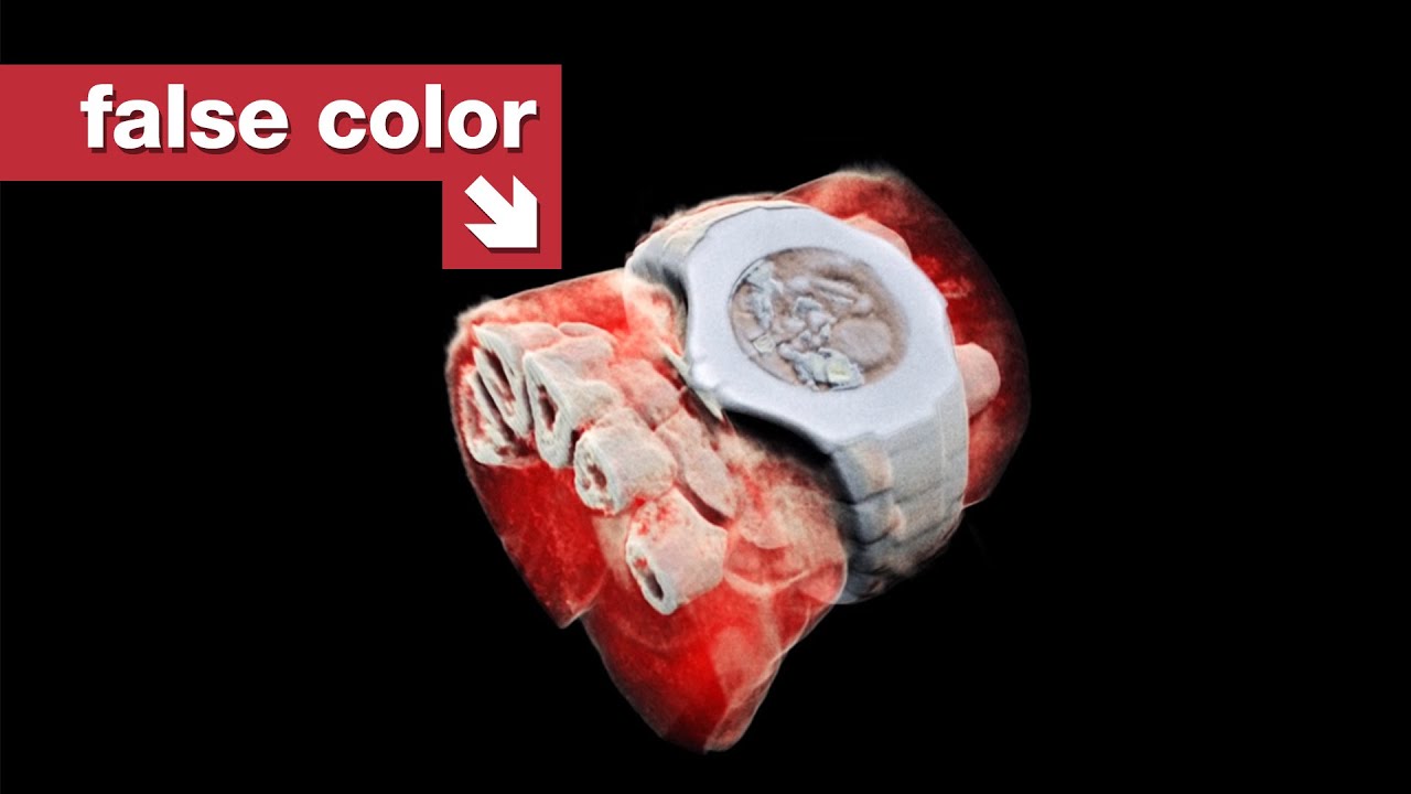
At the University of Canterbury, in Christchurch, New Zealand, the team at Mars Bioimaging are using detector equipment originally developed for the Large Hadron Collider, and putting it to a very different use: medical imaging that allows 3D, false-color images inside the human body.
Thanks to the Mars Bioimaging team: https://www.marsbioimaging.com/
Edited by Michelle Martin https://www.youtube.com/@OnTheCrux
Audio mix by Graham Haerther https://haerther.net/
🟥 MORE FROM TOM: https://www.tomscott.com/
(you can find contact details and social links there too)
📰 WEEKLY NEWSLETTER with good stuff from the rest of the internet: https://www.tomscott.com/newsletter/
❓ LATERAL, free weekly podcast: https://lateralcast.com/ https://youtube.com/lateralcast/
➕ TOM SCOTT PLUS: https://youtube.com/tomscottplus
👥 THE TECHNICAL DIFFICULTIES: https://youtube.com/techdif


@TomScottGo
December 16, 2025 at 12:53 am
There's a line in here that goes by very quickly, but to be clear: that little X-ray sensor measures the frequency of each individual photon that hits each pixel. It's an offshoot of tech from the Large Hadron Collider.
@taylorgay1641
December 16, 2025 at 12:53 am
I'd love to see an update on how this technology has progressed over the past few years
@ohtrueyeahnah
December 16, 2025 at 12:53 am
This is what Capsule Corp was using on Dragon Ball Z! Wow
@adrianadrian2735
December 16, 2025 at 12:53 am
I think that storing parts of the scan on a machine made to store the data on a large number of high transfer rate RAM sticks to be later rendered as a whole by the machine, would be a cheap way of dealing with data storage issues reliably.
In my idea, the scan-data could be quickly stored in and spread between many high transfer rate RAM modules on a machine or a series of machines, the machine then would take the data saved on the modules and, slowly, over time, save it bit-by-bit within the constraints of a conventional storage device's transfer-rates (such as Hard or Solid State Drives), this minimises the exposure time during scan whilst also retaining the data, as during the short-time of the scan, all the scan-data could be quickly "absorbed" by the much higher transfer rate RAM (kind of like a sponge), allowing it to later be "drained" slowly into a whole on a conventional storage device.
Wishing all the best and hope this helps someone at some point perhaps.
@theopoldthegamer4284
December 16, 2025 at 12:53 am
Ew
@Ynhockey
December 16, 2025 at 12:53 am
What happened since? Is there a follow-up? Thanks!
@RD9_Designs
December 16, 2025 at 12:53 am
That's great to know! Thanks Tom! But I can't imagine the googley eyes give anyone comfort when they just serve to make you think that you are going into it's mouth!
@cz5899
December 16, 2025 at 12:53 am
I wish i could take a pic of my feet , i had a bad accident and i just want to KNOW what is going on .. i can only pray
@polerin
December 16, 2025 at 12:53 am
The machine is running Ubuntu. Noice.
@backpacker9131
December 16, 2025 at 12:53 am
Super interesting. I’m glad I stumbled upon this channel… question though, if MRI’s are the current best way of getting a detailed image of the body for doctors/patients, would this replace MRI’s or be used in conjunction with one another? And how much radiation would be at risk of getting from this compared to what we have currently?
@TonyDAnnunzio
December 16, 2025 at 12:53 am
Christchurch was where that massacre happened
@Deadpool25K78
December 16, 2025 at 12:53 am
Would love an update on this
@primodernious
December 16, 2025 at 12:53 am
had been nice to have a portable fossil scanner in color
@leftheading
December 16, 2025 at 12:53 am
Looking forward to about ten years of this technology only being medically available to Football players.
@WillyLangley
December 16, 2025 at 12:53 am
cool
@olibob203
December 16, 2025 at 12:53 am
this is incredible
@TheRisky9
December 16, 2025 at 12:53 am
I wonder if this will help detecting things like damaged tissue or hematomas. For example, it can help when people claim they were injured in an accident, but we can show that they weren't.
@TheCreeperTrack1
December 16, 2025 at 12:53 am
Frylies
@peanuts2105
December 16, 2025 at 12:53 am
And I thought New Zealand was full of Hobbits and Sheep
@tonyd6853
December 16, 2025 at 12:53 am
What if you froze the person before you throw them in that microwave?
@the.Blue42
December 16, 2025 at 12:53 am
⭐😃👍
Comments are closed.